Experience matters
12,000+
cataracts fixed and counting!
Restore your vision today
Your Vision, Your Life
Cataract Surgery Is Where
the Conversation Begins
At Brazos Eye Surgery, cataract surgery is not the end of your care — it is the start of a partnership to reclaim how you see and how you live. Every technical advantage on this page exists for one reason: to give you back the clarity and confidence that blurry vision has been quietly taking away.
Fellowship-trained expertise and premium lens technology are not luxuries here. They are the tools we use to help Waco patients stop adapting to poor vision and start living fully again.
Where the Conversation Begins
We do not treat cataract surgery as a one-and-done procedure. Your evaluation opens a thoughtful planning process — how you read, drive, work, and move through your day — so every decision that follows serves the life you want to live.
A Genuine Vision Upgrade
Expert lens selection goes far beyond clearing a cloudy cataract. With fellowship-trained guidance and the full premium IOL spectrum, we match technology to your anatomy and your goals — delivering a true lifestyle upgrade, not just sharper distance vision.
Autonomy on Your Own Terms
Declining vision quietly reshapes daily life — leaning on family for rides, postponing errands, narrowing where you feel comfortable going. Restored sight returns the independence to drive, travel, and handle your own schedule without asking someone else to get you there.
Full Restoration, Not Partial Fix
Removing the cataract is only the first step. Our goal is to fully restore functional vision — near, intermediate, and distance, as your eyes allow — so you are not trading one limitation for another. Independence means seeing clearly across every distance that matters to you.
See the Procedure
Related
Femtosecond Laser-Assisted Cataract Surgery (FLACS)
When combined with bi-manual technique, femtosecond laser pre-fragmentation reduces phaco energy further — and provides laser-precision capsulotomy for optimal IOL centration. Learn how the two work together.
Why Training Matters
FWCRS-Certified Refractive Cataract Surgeon
vs. Standard Ophthalmology
Every ophthalmologist completes a residency that includes cataract surgery training. But residency is a broad foundation — not subspecialty education in refractive outcomes. Dr. Swann is fellowship-trained and certified by FWCRS — Fellow of the World College of Refractive Surgery & Visual Sciences — meaning his practice is built around refractive cataract surgery: treating the cataract and optimizing the vision you will live with afterward, not simply removing a cloudy lens.
That distinction shows up in surgical technique, diagnostic depth, complication avoidance, and the full range of lens and technology options offered to every patient. A refractive cataract surgeon and a standard ophthalmologist may both perform cataract surgery — but they are not practicing the same level of care.
| Area | FWCRS-Certified Refractive Cataract Surgeon | Standard Ophthalmology |
|---|---|---|
| Training & Certification | ✓ Fellowship-trained refractive cataract surgeon, certified by FWCRS (Fellow of the World College of Refractive Surgery & Visual Sciences) — 1–3 additional years focused exclusively on refractive surgery, cornea, and anterior segment after full ophthalmology residency. | Standard ophthalmology residency includes basic cataract training at lower volume and complexity. No subspecialty fellowship. No FWCRS certification. Most ophthalmologists in general practice perform routine cataracts only. |
| Surgical Approach | ✓ Refractive cataract surgery mindset — bi-manual microincision phacoemulsification, precision astigmatism control, and lens selection designed to optimize final vision, not just remove the cataract. | Standard cataract removal — typically coaxial phacoemulsification with a single, larger incision. Effective for routine cases, but less emphasis on refractive outcome optimization and corneal preservation. |
| Pre-Op Diagnostics | ✓ Full corneal topography, specular microscopy (endothelial cell count), advanced biometry, macular OCT, and Fuchs dystrophy screening on every patient. Refractive planning starts before surgery is scheduled. | Standard biometry for IOL power calculation. Specular microscopy and corneal topography are often omitted unless specifically indicated. Fuchs screening is inconsistent. |
| IOL Portfolio | ✓ Access to and deep experience with the full premium IOL spectrum — monofocal, toric, EDOF, trifocal, light-adjustable, and investigational trial lenses. Recommendation based on refractive goals and anatomy, not surgeon familiarity. | Typically 2–3 IOL options offered. Lens recommendation often defaults to the lenses the surgeon uses most frequently — not necessarily the best refractive match for the patient's anatomy and lifestyle goals. |
| Complex Case Management | ✓ Trained and experienced in small pupils, pseudoexfoliation, zonular instability, prior refractive surgery, dense cataracts, and combined cataract-cornea cases — refractive planning included from the start. | Routine cases handled confidently. Complex anatomy, prior LASIK/PRK, or co-existing corneal disease may require referral to a subspecialist — sometimes after complications have already occurred. |
| Technology Access | ✓ Center of Excellence designation, clinical trial participation, femtosecond laser integration. Early access to refractive technologies before general market release. | Standard phacoemulsification platform. Femtosecond laser may not be available. No trial IOL access. Technology choices limited to what a general ophthalmology practice typically offers. |
Size matters.....
Dr. Swann's Size Instruments
All Other Waco Ophthalmologists
which one do you want in your eye?
Surgical Technique
Bi-Manual Microincision Phacoemulsification
Dr. Swann performs bi-manual microincision phacoemulsification (MICS) — a technique that requires specific fellowship training and is not used by most general ophthalmologists. It is, by every objective measure, the most tissue-sparing approach to cataract removal currently available.
The difference between bi-manual and standard coaxial phacoemulsification is not a matter of surgeon preference. It is a matter of surgical engineering — and the outcomes data support the bi-manual approach, particularly for corneal health and reduced long-term complication risk.
Two Micro-Incisions Under 2mm Each
Bi-manual phaco uses two separate sub-2mm incisions rather than one larger coaxial incision. Smaller openings mean less disruption to the corneal architecture, lower induced astigmatism, and faster wound closure.
Lower Cumulative Phaco Energy
Because the irrigation and aspiration functions are separated into two dedicated instruments, the phaco probe can operate more efficiently with less heat generation. Total ultrasound energy delivered to the eye is measurably reduced — a direct benefit to corneal endothelial cells.
Better Fluid Dynamics Inside the Eye
Separating irrigation from the phaco handpiece allows the surgeon to direct fluid flow with greater precision, maintaining a more stable anterior chamber and reducing the mechanical turbulence that stresses the corneal endothelium.
Faster Healing, Less Induced Astigmatism
Smaller wounds self-seal more reliably, reduce the risk of wound-related astigmatism, and recover faster than larger coaxial incisions. For patients receiving premium IOLs — especially toric and multifocal lenses — this precision matters enormously for final visual outcomes.
Incision size — vs. 2.2–3mm in standard coaxial
Lower cumulative phaco energy delivered to the eye
Smaller wounds induce less surgically-created astigmatism
Better endothelial cell preservation at 1, 3, 6 months post-op
The Missed Diagnosis
Fuchs Dystrophy & Corneal Disease:
You Can Miss What You Don't Know About
Industry-standard cataract workups often stop at biometry — enough to pick a lens power, not enough to see what the cornea is hiding. Hidden corneal disease is one of the most consequential diagnoses a pre-op evaluation can miss. At Brazos Eye Surgery, advanced screening is part of every cataract evaluation — not an optional upsell.
What Is Fuchs Endothelial Dystrophy?
Fuchs dystrophy is a progressive disease of the corneal endothelium — the single-cell inner layer responsible for keeping the cornea clear by pumping fluid out of the tissue. As endothelial cells deteriorate, the cornea slowly swells, clouds, and eventually becomes painful and visually disabling.
It is far more common than most patients realize — estimated to affect up to 4% of adults over 40 in some populations. Many have it without any symptoms. And many have cataract surgery without anyone checking for it first.
Why It Gets Missed
Diagnosing Fuchs at the pre-cataract stage requires equipment most general ophthalmology offices do not have — specifically a specular microscope (which photographs and counts endothelial cells) and the slit-lamp expertise to identify guttata before the disease becomes visually significant.
Without this screening, a surgeon may perform cataract surgery on a cornea that cannot tolerate the procedure — triggering decompensation that leads directly to a corneal transplant that might have been avoided entirely.
- Cataract surgery on an undiagnosed Fuchs cornea dramatically increases the risk of post-operative corneal decompensation
- Decompensated corneas cause chronic pain, blurred vision, and light sensitivity — often requiring a corneal transplant to resolve
- Without a specular microscope and slit-lamp expertise, Fuchs dystrophy is easily missed on a routine pre-op exam
- Corneal topography and anterior OCT can reveal guttata and endothelial irregularities before any visible clouding occurs
- Early identification allows for surgical planning that minimizes energy exposure — or, in advanced cases, a combined cataract-cornea procedure that avoids two separate surgeries
Our Corneal Screening Protocol
Standard with every cataract evaluation — not an add-on
You can miss what you don't know about — so we screen for it before surgery is ever scheduled. Every patient at Brazos Eye Surgery receives a full corneal workup as part of the standard cataract evaluation: specular microscopy, corneal topography, anterior segment OCT, and a dedicated slit-lamp exam for guttata. We are looking specifically for Fuchs dystrophy, map-dot-fingerprint dystrophy, keratoconus, prior refractive surgery changes, and any condition that would change how we plan your procedure — problems that basic industry workups routinely overlook until after complications appear.
Specular Microscopy
Standard on every evaluation at Brazos — counts and photographs endothelial cells. Industry norm: omitted unless corneal disease is already suspected.
Corneal Topography
Included routinely, not reserved for 'complicated' cases — maps the full corneal surface for irregularities, astigmatism axis, and keratoconus screening.
Anterior Segment OCT
Cross-sectional corneal imaging before surgery — reveals structural abnormalities that basic biometry and a quick slit-lamp glance cannot.
Slit-Lamp Guttata Exam
Direct Fuchs screening by fellowship-trained cornea expertise — not the cursory pre-op check many cataract evaluations stop at.
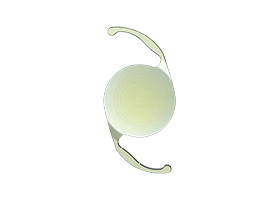
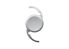
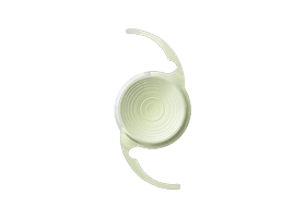
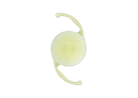
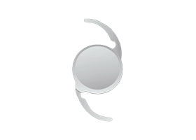
Premium IOL Options
The Full Spectrum of Lens Technology
Cataract surgery is the only procedure in medicine that removes a focusing element of the eye and replaces it with something potentially better. The lens you choose determines your vision for the rest of your life. We carry every category of premium IOL — including trial lenses not yet available at most practices in the country.
As a Center of Excellence and clinical trial evaluation site, Brazos Eye Surgery gives select patients access to next-generation IOL technology before it reaches the general market — ensuring our patients are always at the leading edge of what is possible.


Monofocal
Distance clarity
Toric
Astigmatism correction
EDOF
Distance to intermediate
Trifocal / Multifocal
Full spectacle independence
Light-Adjustable
Adjustable after surgery
Trial IOLs
Pre-release access
Center of Excellence — Clinical Trial Site
Every lens. Explained honestly.
Including the ones not yet released.
Our Advanced Lens Technology Center covers every IOL option in full — what each lens does, how it works, who it is best for, and what the honest trade-offs are. Plus: what it means to be a clinical trial site, and how that gives our patients access to technology years ahead of most practices.
Join Fellow Central Texans Enjoying Worry-Free Vision
Real cataract patient experiences from Central Texans who chose local care with Dr. Swann — not a drive to Austin or Dallas.
Your experience helps others in Waco find the care they deserve. Share your story on Google.
Leave a reviewQuotes are summarized from verified Google reviews and cataract patient testimonials at Brazos Eye. See all reviews on Google · Leave yours
After Cataract Surgery
Waxy or Cloudy Vision After Cataract Surgery?
Vision sharp at first, then slowly hazy again? That is often posterior capsule opacification — sometimes called a secondary cataract. It is common, easy to treat with an in-office YAG laser, and usually not part of your original cataract surgery coverage. Learn what to expect.
Explore Further
FWCRS vs. Standard
What FWCRS-certified refractive cataract training means for your outcome
Bi-Manual Technique
The most tissue-sparing cataract technique available
Femtosecond Laser (FLACS)
Laser precision for capsulotomy, fragmentation, and incisions
Fuchs & Corneal Screening
Why we check what most surgeons skip
Advanced Lens Technology
Every IOL, explained — including pre-release trial lenses
Waxy Vision After Cataract
PCO, insurance coverage, and YAG laser treatment explained
Ready to See Clearly?
Schedule Your Cataract Evaluation
Full corneal workup. Honest lens recommendation. Fellowship-trained surgical expertise — right here in Waco.